|
무당벌레에서 유래된 HaGF peptide를
이용한 염증 억제 효과
김동희2, 김현정1,3, 이진영4, 황재삼5, 김인우5, 이슬기1, 정현국1, 안봉전1*
1대구한의대학교 화장품약리학과, 2한국한방산업진흥원, 3허브누리,
4호서대학교 한방화장품과학과, 5국립농업과학원
|
본 연구에서는 무당벌레 유충으로부터 분리된 항균 펩타이드 유전자의 일부 영역인 HaGF를 이용하여 대식세포의 염증에 미치는 영향을 조사하였다. 세포는 LPS 처리 후 한 시간 뒤에 HaGF를 처리를 하여, 세포 독성이 나타나지 않는 농도인 5, 25, 50, 100 μg/ml을 사용하였다. 그 결과 HaGF가 염증성 cytokine의 생성을 감소시키는 것을 확인 하였으며 iNOS와 COX-2 역시 100 μg/ml의 농도에서 각각 51%, 49% 저해율을 보였다. 따라서, HaGF는 LPS로 유도된 대식세포주인 Raw 264.7 세포에서의 염증 반응 억제 효과를 기대할 수 있었다.
|
서 론
예로부터 곤충은 동양에서 약용?식용으로 많이 이용되었는데, 고서의 기록에 의하면 동의보감은 약 95종의 약용 곤충이, 본초강목습유(本草綱目拾遺)에는 106종의 약용 곤충이 기록되어있다[22]. 이러한 곤충자원은 최근 생명공학 기술의 발달과 함께 다양화 되고 있는데, 그 예로 각종 유용물질을 생산하는 누에(실크, 누에가루, 동충하초), 꿀벌(꿀, 로얄제리. 프로폴리스), 곤충의 외골격(chitin의 추출) 등이 좋은 예라 할 수 있다. 또한 곤충의 추출물은 전통적인 생약으로 이용되었는데, 최근 이러한 곤충 추출물에 대한 효능 및 유효성분 분석에 대한 연구가 진행되고 있다. 이 외에도 곤충 자원은 유용생물자원으로 현재 세계적으로 여러 분야에서 매우 광범위하게 개발 활용되고 있다[6, 7, 8, 12, 16].
염증은 생체 혹은 조직에 물리적 자극이나 화학적 물질, 세균 감염 등의 기질적 변화를 가져오는 침습이 가해질 때 그 손상부위를 수복 재생하려는 복구기전으로서 자극이 가해지면 국소적으로 염증성 성분이 증가되면서 염증이 유발된다[14]. 임상적으로는 발적, 발열, 종창, 동통, 기능장애 등의 증상이 나타난다[11]. 대식세포는 선천면역뿐만 아니라 획득면역 등 다양한 숙주반응에 관여하여 항상성 유지에 관여하는 것으로 알려져 있으며, 염증반응 시에는 nitric oxide (NO)와 cytokine을 생산하여 감염초기에 생체방어에 중요한 역할을 한다[3]. 포유동물 세포의 nitric oxide synthase (NOS)의 경우, 유사형태가 3가지 존재하는데 neuronal NOS (nNOS), endothelial NOS (eNOS), 그리고 inducible NOS (iNOS)이다. 그 중에서 특히 iNOS가 염증반응에 관여한다. nNOS와 eNOS는 항상 발현되어 있으며, iNOS의 경우 interferon-γ, lipopolysaccharide (LPS), 그리고 여러 가지 염증 cytokine의 자극이 있을 때 발현된다. 또한 NO는 급성, 만성 염증반응을 조절하기도 한다. COX에 대해서는 1990년대 초반에 주로 연구되었는데, 이 또한 유사형태가 두 가지 존재한다. COX-1은 거의 모든 조직에 발현되어 있고, prostagrandin을 생산하여 신장의 혈액흐름을 조절하거나 위장의 세포를 보호하는 등의 생리적인 기능을 조절한다. 반대로 COX-2의 경우는 미생물에 의한 감염이나 손상 혹은 여러 요인의 스트레스에 반응한 대식세포 (macrophage)에서 발현된다. 즉 iNOS와 COX-2의 발현유무는 면역 세포의 염증 반응에 관여한다. 또한 염증 사이토카인(proinflammatory cytokines)인 tumor necrosis factor-α (TNF-α), interleukin-1β (IL-1β), interleukin-6 (IL-6), monocyte chemoattractant protein-1 (MCP-1)등이 염증 반응에 관여하는 인자로 알려져 있다[9, 24].
따라서 본 연구에서는 무당벌레 유충으로부터 분리된 펩타이드 유전자 Harmoniasin의 일부 영역 HaGF을 이용하여 LPS로 활성 된 Raw 264.7 cell로 염증에 관련된 다양한 기전과 단백질 발현을 측정하였다
본 실험에서 사용한 펩타이드 HaGF는 AnyGen (Gwang-ju, Korea)사에서 98% 이상의 순도로 합성하였고, 0.01% acetic acid에 녹여 사용하였다
결과 및 고찰
Raw 264.7 cell에 대한 독성
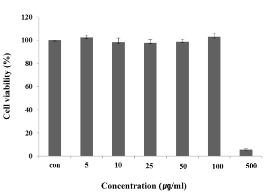
마우스 대식세포인 Raw 264.7 cell에 대한 HaGF의 세포독성을 확인하기 위하여 MTT assay를 수행하였다. HaGF를 농도 별로 24시간 동안 처리한 결과 100 μg/ml의 농도까지는 독성이 나타나지 않았지만, 500 μg/ml의 농도에서는 세포의 생존율을 95% 감소시켰다(Fig. 1). HaGF는 100 μg/ml 이하의 농도에서는 세포독성이 낮아 세포의 생존율에 영향을 주지않는다는 사실을 확인할 수 있었다.
Fig. 1. Cell viability of HaGF on Raw 264.7 cell. Raw 264.7 cells were treated with 5, 10, 25, 50, 100, 500 μg/ml of HaGF dissolved in media for 1 hr prior to the addition of LPS(1 μg/ml), and the cells were further incubated for 24hr. Data represent the mean±S.D. with eight separate experiments.
Nitric oxide (NO) 생성억제 효과
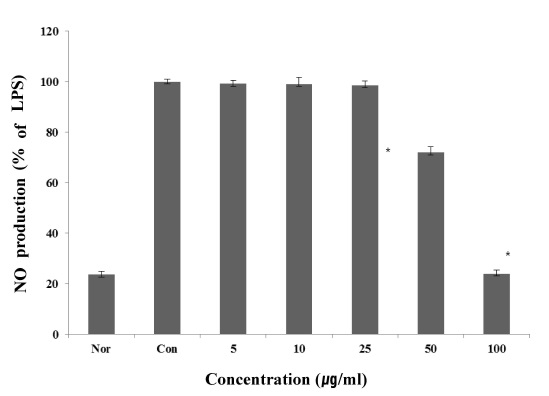
NO는 박테리아를 죽이거나 종양을 제거시키는 역할을 하지만, 병리적 원인에 의해 과도하게 생성, 분비되면 염증을 유발시키게 되며, 조직 손상, 유전자 변이 및 신경손상 등을 일으키는 것으로 알려져 있다[13, 20]. 또한, NO는 NO 합성효소에 의해 l-arginine으로부터 생성되는 무기 유리체로 면역반응, 세포독성, 신경 전달계 및 혈관이완 등 여러 가지 생물학적인 과정에 관여하는 것으로 알려져 있으며, 농도에 따라 세포기능유지에 중요한 작용을 하기도 하고 세포독성을 일으키기도 한다[14, 17]. NO는 그 자체로 염증유발과 조직손상을 일으킬 뿐만 아니라, 활성산소인 superoxide와 결합하게 되면 보다 반응성이 강한 peroxynitrite의 형태로 전환되어 생체조직의 괴사를 야기할 수 있다[19]. 이에 본 실험에서는 NO 생성에 대한 HaGF의 효과를 알아보았다. 생성된 NO양을 griess 시약을 이용하여 세포배양액 중에 존재하는 NO2-의 형태로 측정하여 Fig. 2에 나타내었다. 그 결과 LPS 처리 후 NO 생성량은 정상세포에 비하여 약 4배 이상 증가되었다. HaGF를 100 μg/ml의 농도로 처리한 군에서는 LPS로 증가된 NO 생성량이LPS 처리군에 비하여 50% 이상 감소함을 확인할 수 있었다.
Fig. 2. Inhibitory effects of HaGF on the production of nitricoxide Raw 264.7 cells. Raw 264.7 cells were cultured with LPS (1 μg/ml) in the presence or absence of HaGF for 24 hr to determine the level of NO. (Nor: LPS not induced group, Con: LPS induced group) The data represent the mean±SD of three separate experiments(significant as compared to control *p<0.05).
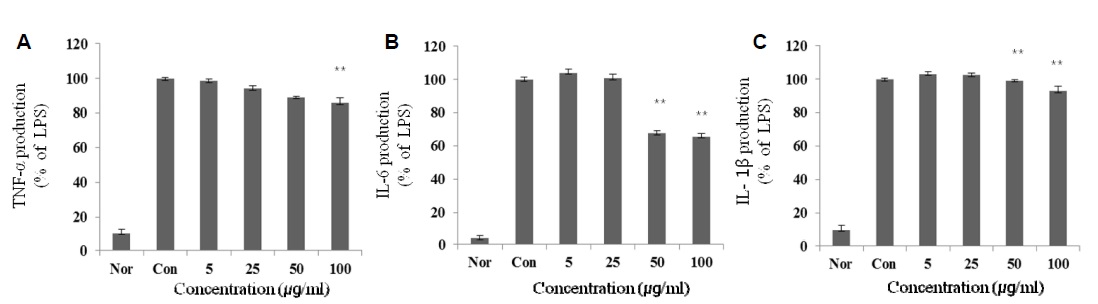
TNF-α, IL-1β, IL-6 생성 억제 효과
TNF-α를 포함한 매개물질인 cytokine IL-6와 IL-1β는 NF-κB를 통해 활성화 되고 또한 NF-κB를 활성화 시켜서 cytokinecascade를 증폭하고 염증상태를 확장한다[2]. 특히, TNF- α는 염증반응에 있어서 중요한 역할을 하며 macrophage와 mastcell 등에서 분비되며, LPS반응의 주요 매개체로서 내재면역에 있어서도 중요한 역할을 하며 만성 염증 반응과도 관련되어 있다[10]. IL-1β는 T-cell의 활성화, B-cell의 성숙, NK cell의 activity를 활성화 하며, IL-6는 림프구를 활성화시켜 항체생산을 증가시키는 것으로, IL-6의 level은 염증 반응에서 항상 증가하는 것으로 보고되고 있다[1]. 본 실험에서 LPS는 TNF-α, IL-1β, IL-6의 생성을 증가 시켰지만, HaGF를 농도 별로 처리한 군에서는 억제되는 것을 확인할 수 있었다(Fig. 3).
Fig. 3. Effect of HaGF on the production of cytokines stimulated by LPS. Production of TNF-α (a), IL-6 (b), IL-1β (c) were measured in the medium of Raw 264.7 cells cultured with LPS (1 μg/ml) in the presence or absence of HaGF for 24 hr. The amount of TNF-α was measured by immunoassay as described in materials and methods. (Nor : LPS not induced group, Con: LPS induced group) Data represent the mean±S.D. with three separate experiments. One-way ANOVA was used for comparisons of multiple group means followed by t-test (significant as compared to control *p<0.05, **p<0.01).
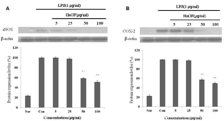
iNOS, COX-2의 단백질 발현 저해 효과
iNOS는 평소에는 세포 내에 존재하지 않으나 일단 유도되면 장시간 동안 다량의 NO를 생성하며, 생성된 NO는 혈관투과성, 부종 등의 염증 반응을 촉진 시킬 뿐만 아니라 염증매개체의 생합성을 촉진하여 염증을 심화시키는 것으로 알려져 있다[5, 21]. 염증반응과 관련된 조직 손상에서 NO와 iNOS의 발현이 증가되어 있음이 보고된 바 있다[15, 18, 23] COX-2는 cyclooxygenase와 peroxidase 활성을 모두 가지고 있는 효소이다. Cyclooxygenase 기능으로서 arachidonic acid를 prostagland로 변환하고, peroxidase 기능으로서는 endoperoxide를 prostaglandin로 변환시키며, prostaglanin는 prostaglandins,thromboxane 및 prostacyclins의 전구체로 사용된다. COX-1은 모든 세포에 존재하면서, 정상세포의 항상성을 유지하며, COX-2는 급성염증반응에서 prostaglandin의 합성에 관여하고, LPS 및 cytokine에 의해 발현이 유도된다[4, 9]
Fig. 4. Inhibitory effects of HaGF on the protein levels of iNOS and COX-2 in Raw 264.7 cells. Raw 264.7 cells (5x10⁵ cells/ml) were pre-incubated for 24 hr, and the cells were stimulated with lipopolysaccharide (1 μg/ml) in the presence of complex extracts sample (5, 25, 50, 100 μg/ml) for 24 hr. (Nor: LPS not induced group, Con: LPS induced group) Data represent the mean±S.D. with three separate experiments. One-way ANOVA was used for comparisons of multiple group means followed by t-test (significant as compared to control *p<0.05, **p<0.01).
감사의 글
본 논문은 농촌진흥청 차세대 바이오그린21사업(과제번호:PJ008158)의 지원에 의해 이루어진 것임.
References
1. Delgado, A. V., McManus, A. T. and Chambers, J. P. 2003.
Production of tumor necrosis factor-alpha, interleukin 1-beta,
interleukin 2, and interleukin 6 by rat leukocyte subpopulations
after exposure to substance. P Neuro 37,
355-361.
2. Hanada, T. and Yoshimura, A. 2002. Regulation of cytokine
signaling and inflammation. Cytokine Growth Factor Rev 13,
412-421.
3. Higuchi, M., Hisgahi, N., Taki, H. and Osawa, T. 1990.
Cytolytic mechanisms of activated macrophages. Tumor necrosis
factor and L-arginine-dependent mechanisms act synergistically
as the major cytolytic mechanisms of activated
macrophages. J Immunol 144, 1425-1431.
4. Hume, D. A., Wells, C. A. and Ravasi, T. 2007.
Transcriptional regulatory networks in macrophages.
Novartis Found Symp 281, 2-18.
5. Kim, R. G., Shin, K. M., Chun, S. K., Ji, S. Y., Seo, S. H.,
Park, H. J., Choi, J. W. and Lee, K. T. 2002. In vitro anti-inflammatory
activity of the essential oil from ligularia fischeri
var. spiciformis in murine macrophage RAW 264.7 cells.
Yakhak Hoeji 46, 343-347.
6. Kou, J., Ni, Y., Li, N., Wang, J., Liu, L. and Jiang, Z. H.
2005. Analgesic and anti-inflammatory activities of total extract
and individual fractions of Chinese medicinal ants
Polyachis lamellidens. Biol Pharm Bull 28, 176-180.
7. Kurioka, A. and Uamazaki, M. 2002. Purification and identification
of flavonoids from the yellow green cocoon shell
(Sasamayu) of the silkworm, Bombys mori. Biosci Biotechnol
Biochem 66, 1396-1399.
8. Li, N. G., Osakovskii, V. L. and Ivanova, S. S. 2003. Chemical
composition and cryoprotective activity of ethanol extract
from winter caterpillars Aporia crataegi L. Izv Akad Nauk Ser
Biol 5, 547-552.
9. Lin, W. J. and Yeh, W. C. 2005. Implication of Toll-like receptor
and tumor necrosis factor alpha signaling in septic
shock. Shock 24, 206-209.
10. Lee, A. K., Sung, S. H., Kim, Y. C. and Kim, S. G. 2003.
Inhibition of lipopolysaccharide -inducible nitric oxide synthase,
TNF-α and COX-2 expression by sauchinone effects
on I-κBα phosphorylation, C/EBP and AP-1 activation.
British J Pharmacol 139, 11-20.
11. Lee, T. H., Kwak, H. B., Kim, H. H., Lee, Z. H., Chung,
D. K., Baek, N. i. and Kim, J. 2007. Methanol extracts of
Stewartia Korean inhibition cyclooxygenase-2 (COX-2) and
inducible nitric oxide synthase (iNOS) gene expression by
blocking NF-Kappa B transactivation in LPS-activated Raw
264.7 cells. Mol Cells 23, 398-404.
12. Ma, Y., Wang, X., Zhao, Y., Kawabata, T. and Okada, S.
1997. Inhibitory effects of Chinese and extract (CAE) on
nephrotoxycity induced by ferric-nitrilotriacetate (Fe-NTA)
in Wistar rats. Res. Commun Mol Pathol Pharmacol 96,
169-178.
13. Marin, J. and Rodriquez-Martinez, M. A. 1997. Role of vascular
nitric oxide in physiological and pathological
conditions. Pharmacol Ther 75, 111-134.
14. Moncada, S., Palmer, R. M. and Higgs, E. A. 1991. Nitric
oxide: physiology, pathophysiology and pharmacology.
Pharmacol Rev 43, 109-142.
15. Mori, M. 2007. Regulation of nitric oxide synthesis and apoptosis
by arginase and arginine recycling. J Nutr 137,
1616-1620.
16. Nakatani, T., Konishi, T., Miyahara, K. and Noda, N. 2004.
Three novel cantharidin-related compounds from the
Chinese blister beetle, Mylabris Phalerata Pall. Chem Pharm
Bull 52, 807-809.
17. Nathan, C. and Xie, Q. W. 1994. Nitric oxide synthases: roles,
tolls and controls. Cell 78, 915-918.
18. Palmer, R. M., Ashton, D. S. and Moncada, S. 1988. Vascular
endothelial cells synthesize nitric oxide from L-arginine.
Nature 333, 664-666.
19. Radi, R., Beckman, J. S., Bush, K. M. and Freema, B. A. 1991.
Peroxynitrite oxidation of sulfhydryls the cytotoxic potential
of superoxide and nitric oxide. J Biol Chem 266, 4244-4250.
20. Rodeberg, D. A., Chaet, M. S., Bass, R. C., Arkoviz, M. S.
and Garcia, V. F. 1995. Nitric oxide: An overview. American
J Surgery 170, 292-303.
21. Tezuka, Y., Irikawa, S., Kaneko, T., Banskota, A. H.,
Nagaoka, T., Xiong, Q., Hase, K. and Kadota, S. 2001.
Screening of Chinese herbal drug extracts for inhibitory activity
on nitric oxide production and identification of an active
compound of zanthoxylum bμgeanum. J Ethnopharmacol
77, 209-217.
22. Wang, C. and Leger, R. J. 2005. Developmental and transcriptional
responses to host and nonhost cuticles by the
specific locust pathogen Metarhizium anisoplae var.
acridum. Eukaryot Cell 4, 937-947.
23. Weisz, A., Cicatiello, L. and Esumi, H. 1996. Regulation of
the mouse inducible-type nitric oxide synthase gene promoter
by interferon-γ, bacterial lipopolysaccharide and
NG-monomethyl-L-arginine. Biochem J 316, 209-215.
24. Yun, H. J., Heo, S. K., Yi, H. S., Kim, C. H., Kim, B. W.
and Park, S. D. 2008. Anti-inflammatory effect of injinho-
tang in RAW264.7 Cells. Korean J Herbol 23, 169-178.
논문 전문 바로보기
|